What is MRI?
Siemens 3 Tesla (MRI)
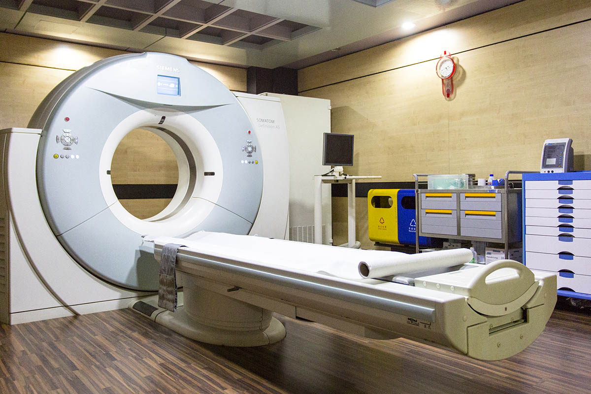
The department is equipped with SIEMENS Somatom definition AS+, 128 slice Cardiac CT & Syngo Via. It is a single source applying lowest radiation dose possible with faster rotation time. The scanner is capable of adapting to virtually every patient and every clinical question. Fueled by the FAST CARE platform, the SOMATOM Definition AS+ is designed to help maximize clinical outcome and raise patient-centric productivity.
The high end CT covers the whole body in seconds. Because of increased speed and resolution the CT produces excellent images of coronary arteries. In addition high resolution studies of Cerebral, Carotid & Vertebral, Pulmonary Artery, Aorta, Renal, Upper Limb, Lower Limb, Abdominal Arteries, Portal Venography, Urogram and CT Enteroclysis are demonstrated as part of the protocol. The latest upgraded software Syngo Via gives excellent reconstruction like Multiplanar Reconstruction, MPI and VRT with our equipment.
Other studies like CT Brian, Orbit, PNS, Neck, Thorax, Abdomen, Spine, Extremities, High Resolution CT of Lung, Temporal bone & joints are routinely done.
The CT scan are also used for procedures like guided fine needle aspiration cytology and biopsies from masses in Thorax, Abdomen, Spine and extremities. CT guided aspiration and drainage tube placement for abscess is done as well.
Siemens 1.5 Tesla (MRI)
The department is equipped with SIEMENS Magnetom Avanto 1.5 Tesla 18 Channels Supercon MRI (Software Version: MR- B 17) a noninvasive imaging that helps Physicians diagnose and treat diseases.
MR imaging uses a powerful magnetic field, radio frequency (63 MHz) pulses and advanced computing to produce detailed images of organs, soft tissues, bone and virtually all other internal body structures. The signals are reconstructed by high end processors this reconstruction also can be made into three-dimensional images, allowing complete and remarkable visualization of the anatomy scanned from various angles. The images are studied by skilled radiologists on the computer monitor and then printed or copied to CD.
Detailed MR images allow physicians to better evaluate parts of the body and certain diseases that may not be assessed adequately with other imaging methods such as X-ray, ultrasound or computed tomography (also called CT or CAT scanning).
MR Mammography, SVS (Single Voxel Spectroscopy) and Cardiac MRI are the recent advantage of Magnetom Avanto 1.5 Tesla 18 Channels Supercon MRI.
The high end MRI System Cerebral, Carotid & Vertebral arteries are demonstrated. Other studies like Neurology, Orthopedics, Body Imaging, Prostate MRI, Pediatric MRI, Oncology and MR Angiography are routinely done.
Limitations of MRI
MRI are still contraindicated in those circumstances- Patient having Cardiac pacemaker, Metallic implants, Aneurysm, Coronary Stent (Magnetic) and Pregnancy (4 months of pregnancy- No known harm but safety of infant from radio-frequency).
Contrast Media
Certain types of MRI scans require the use of a safe para-magnetic agent or contrast media. This is to enhance the blood vessels or certain body parts for the accuracy of your imaging test. The Food & Drug Administration (FDA) has requested manufacturers of gadolinium based contrast add additional warnings to their product labels. To read the FDA warnings regarding gadolinium contrast agents visit their website and view “The Information for Healthcare Professionals” document. You should receive complete instructions concerning the use of a contrast agent from your physician when he or she orders the test. Tell your physician before your exam if you have ever had an adverse reaction to contrast media.
Types of MRI
- Angiography (MRA) – Vascular anatomy visualized
- Breast MRI
- Cardiac MRI
- MRCP – MRI of hepatobiliary and pancreatic system
- Neurology/Central Nervous System
- Orthopedic
- Spectroscopy
Conditions Diagnosed by MRI
- Bone Cancer
- Brain Cancer
- Head & Neck Cancer
- Kidney Cancer
- Liver Cancer
- Pancreatic Cancer
- Stomach Cancer
- Uterine Fibroids
Preparing for your MRI
In general, there are no special preparations to follow before your exam. Because MRI uses a strong magnetic field, metal objects may interfere with the scan. For your convenience, we provide a place to store your keys, jewelry and other valuables during the exam. We ask that you wear comfortable, loose-fitting clothing, but you may be asked to change into a hospital gown for image quality and safety reasons.
Please check the following list carefully. All metallic/surgical implants must be assessed for safety before undergoing any MRI procedure. Common implants that may not be safe for MRI procedures include the following:
- Pacemaker
- Aneurysm clips in the brain
- Inner ear (cochlear) implants
- Implanted spinal cord stimulator
- Metallic implants
- Metal fragments in one or both eyes
Also, please alert our staff if you:
- Have dental bridges
- Wear a hearing aid(s)
- Have ever been a metal worker
- Are pregnant or think you might be
Because a paramagnetic agent (a type of contrast media) may be used, please tell your physician if you:
- Are pregnant or think you might be
- Are breastfeeding
- Have anemia or any diseases that affect red blood cells
- Have asthma or other allergic respiratory disorders
MRI FAQ
No. Since MRI is “non-invasive,” the exam is painless. However, your doctor may utilize a contrast agent to better visualize a part of your anatomy. If this is the case, you may receive a simple injection during the exam.
MRI allows doctors to see images of your internal organs and structures in great detail and from multiple planes. This gives them information quickly and in many cases more accurately than tests used in the past or exploratory surgeries.
No, but you will hear a loud knocking or buzzing sound at various intervals throughout your exam. Other than that, you won’t feel a thing. Ear plugs are available to you for your exam and their use will not prevent you from hearing the technologist if he or she speaks to you during the exam.
No. MRI uses a powerful magnet in conjunction with radio-frequency waves to generate images of your internal organs and structures. There is no ionizing (X-Ray) radiation.
There are very few patients who cannot be comfortably accommodated for an MRI exam. Our equipment has one of the largest openings in the industry at nearly two feet wide.
That will depend on your height and what part of your body is being scanned. The part that is being imaged needs to be in the middle of the magnet. For example, if your ankle is being scanned, your head will be outside of the MRI scanner. If it is your head, neck, or chest being scanned, your head will be inside of the scanner. The new Espree system offers one of the largest and shortest bores in the market helping the claustrophobic patient. With this larger bore design we have the opportunity to keep the patient’s head out of the magnet 60% of the time with certain examinations.
Most people do not experience such a reaction. However, if you have had claustrophobic reactions to enclosed spaces before, you should let the technologist know. Even if you are uncomfortable in small spaces, staff members can help you comfortably complete the study. The Expree system is the closest thing to an Open MRI, offering a 1.5 Tesla high field strength magnet; something Open MRI architecture cannot offer.
You will be in contact with a technologist at all times. Even when he or she is not in the MRI room, you will be able to talk to him or her by intercom. The technologist is always able to see you through a large patient viewing window. In some cases a friend or family member may stay in the scan room with you during your exam, eligibility can be determined during the screening. Please consult the MemorialCare Imaging Center for more information.
The magnet makes a knocking sound as images are being taken. In between scans the machine is quiet. Ear plugs are available to you for your exam and their use will not prevent you from hearing the technologist if he or she speaks to you during the exam.
You do have to remain as still as possible, but the time passes quickly. With new MRI scanners software allows for faster exam acquisition times, reducing the amount of time needed to take the scan. Moving during the procedure may require repeating parts of the exam so it is best to try to remain as still as possible for the best exam results. The amount of time needed for the exam is dependant on what is being studied. A typical exam lasts between 30 and 60 minutes. You should always allow extra time in case the exam lasts longer than expected.
Some patients with metal implants cannot be safely scanned in the MRI environment. People with pacemakers, aneurysm clips (especially in the brain), and neurostimulators generally cannot be scanned. Anyone with surgical pins, shrapnel, plates or other types of metal implants should notify the technologist. You will be required to provide a health history when you arrive for your exam that explains any metallic implants you may have. A doctor will determine if a particular metal implant is approved to be in an MRI environment.