What is Ultrasonographyn?
How Ultrasound Works
Booking and Preparation
You will need to make an appointment for this examination. Depending on the area of the body to be examined you may be asked to fast from food and fluids for a period of time before your scan. Alternatively, some examinations require you to drink a significant amount of water prior to arriving so that your bladder is full. You will be advised of the appropriate preparation when you make your appointment.
Ultrasound Technologists
Our panel of Ultrasound Technologists and Patient Care officers exceeds 40 personnel. We are here to ensure a comfortable experience for the patient during the diagnosis followed by a swift accurate result for diagnosis purposes.
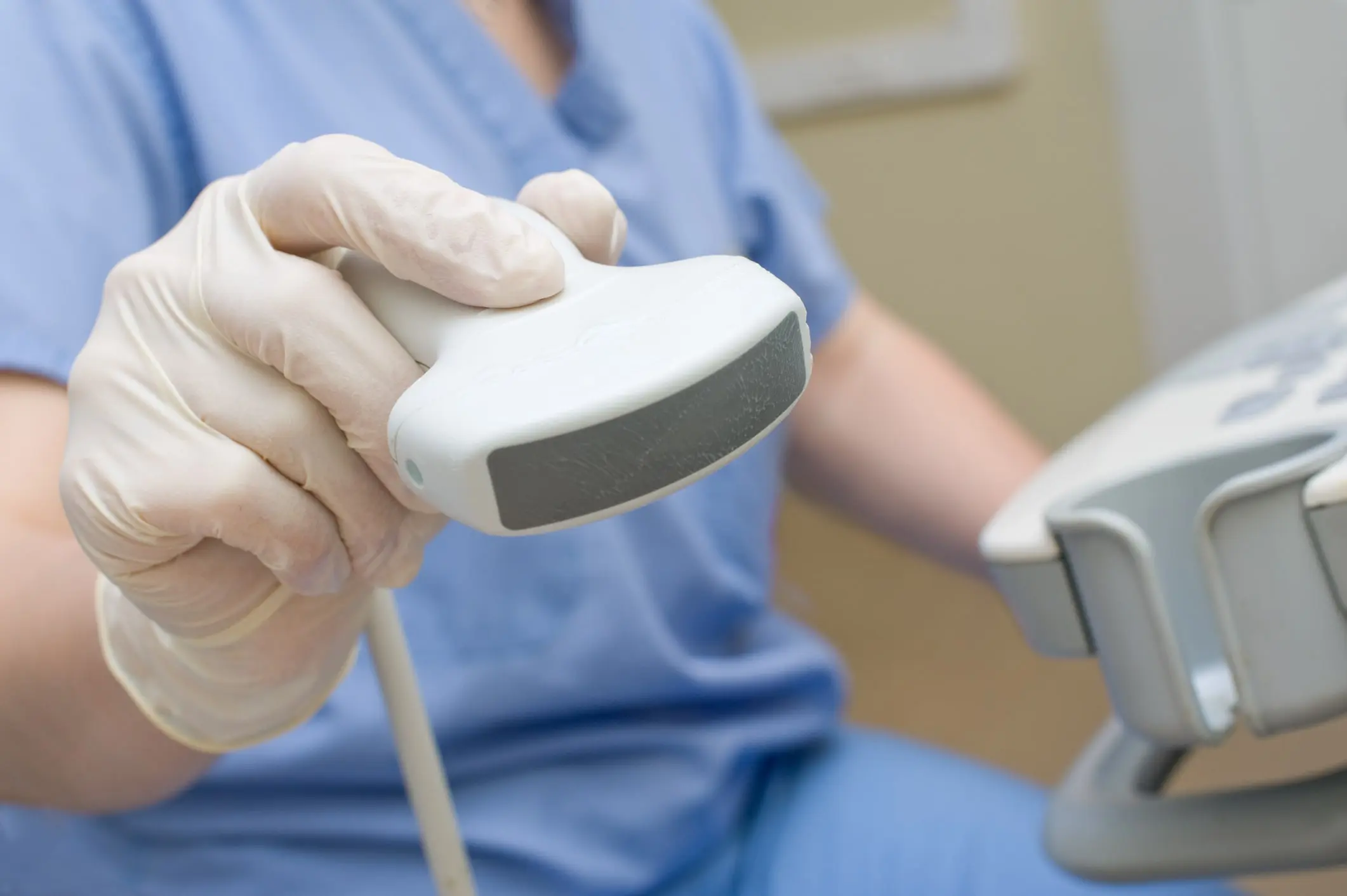
Who will perform the scan?
Your examination or scan will be performed by either a Radiologist (a highly trained specialist doctor ) or a sonographer (a specially trained technologist). Because the examiner is interpreting moving images on a screen a high degree of concentration is required to obtain accurate information. Therefore, in most circumstances except obstetric scans, family and friends of the patient are not generally permitted to watch the procedure. If you have accompanying children you will have to bring along someone to watch them during your examination.
Types of Ultrasound
Different types of ultrasound are offered at specific imaging center locations, some include:
- General Ultrasound
- Abdomen
- Pelvis
- Prostate
- Testicular
- Thyroid
- Breast Ultrasound
- Obstetrical Ultrasound
- Pediatric Ultrasound
- Transrectal Ultrasound
- Transvaginal Ultrasound
- Ultrasound-Guided Biopsy
- Ultrasound-Guided Paracentisis
- Ultrasound-Guided Thoracentesis
- Vascular Studies for Veins and Arteries
- Abdominal Ultrasound
- Ankle-Brachial Index (ABI)
- Arterial Duplex Scan
- Carotid Ultrasound
- Venous Duplex Scan
Procedure
For most ultrasound examinations you will be required to change into a gown and lie on an examination couch. You will remain covered during your examination except for the area being imaged. In order to obtain optimal images a layer of gel will be applied to the area being imaged so that good contact is made between you and the ultrasound probe. The probe will be placed directly onto the gel and your skin for the duration of the examination. Ultrasound examinations are not painful and generally are not invasive but sometimes they can be uncomfortable if you have to maintain a full bladder or move a body part that might be causing you some discomfort e.g. a shoulder.
Colour Doppler ultrasound uses a special technology that looks at blood flow through the arteries and veins, for example – the carotid arteries in the neck that supply blood to the brain, or the veins of the legs. If you are having this examination it is common to hear strange noises as the signals coming from the flowing blood are converted into sound. Sometimes the sonographer will have to gently squeeze the calf a few times when examining the veins in the legs. This should not be painful.
Ultrasound Biopsies
On occasion, a biopsy, or sample of tissue, may be required by your doctor to diagnose a medical problem. If you are required to have a liver or breast biopsy, the test may need to be performed at a specific branch of Perth Radiological Clinic. Whilst many biopsies have no specific requirements, biopsies of the kidney and liver require special preparation (you will be advised by our booking staff when you make your appointment). The specimen will then be sent to a pathology laboratory for processing and the results will be forwarded to your doctor.
Ultrasound Guided Injection
Sometimes your referring doctor may request you to have a pain relieving injection into a specific area. A radiologist will perform this procedure and the ultrasound probe is used to guide the injection to the correct place. No specific preparation is required for this procedure.
Conditions Diagnosed by USG
Ultrasound can be used to image many areas of the body including the pelvis and abdomen, the musculoskeletal system, the breast, the male reproductive system, the kidney, the thyroid, and salivary glands, the gall bladder, the pancreas and the developing fetus.
- Bone Cancer
- Gynecologic Cancer
- Kidney Cancer
- Liver Cancer
- Ovarian Cancer
- Pancreatic Cancer
- Prostate Cancer
- Testicular Cancer
- Thyroid Cancer
- Uterine Cancer
- Heart Attack (Myocardial Infarction)
- Heart Disease
- Peripheral Vascular Disease
- Uterine Fibroids
- Varicose Veins
Pregnancy Ultrasound
Pregnancy ultrasounds may be performed by examination of the abdomen called transabdominal or by using a special probe designed to be inserted into the vagina, called transvaginal. The type of examination you will need will depend on what your referring doctor has requested and the nature of the clinical condition being investigated.
Preparation
Early pregnancy scan:
- Finish drinking 750ml of water 1 hour prior to exam
- Do not empty bladder
- Continue to take any current medication
Anatomy pregnancy scan:
- Drink 2 glasses of water half an hour prior to your appointment
- Do not empty bladder
- Continue to take any current medication
Indications for pregnancy ultrasound
Ultrasound is a highly valuable diagnostic tool and it is particularly useful during pregnancy because it is completely safe for you and your baby. Some of the useful indications for use during pregnancy include:
- for dating purposes and to accurately determine your due date
- to ascertain the number of babies present
- to check for any bleeding early in the pregnancy
- to check the position of the placenta
- to assess the growth of the baby and its general well-being
- to provide information about the anatomy of the baby and check for possible abnormalities
How long will the procedure take?
Most ultrasound examinations will be completed within 30 minutes, however, some studies will take longer especially the colour Doppler studies. It is not unusual for the radiologist to come in and speak with you and view the images on the screen. The radiologist will then interpret all images produced during the examination and the results will be forwarded to your doctor.
Please note: that due to the high demand for ultrasound examinations, a cancellation fee will be charged if you do not give at least 24 hours notice for a cancelled appointment.
Please remember to bring any previous ultrasound or x-ray films to your appointment.